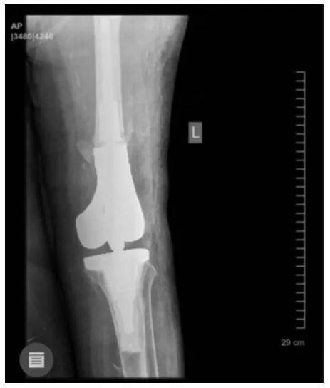
The patient is an 86-year-old woman who presented to the doctor with a periprosthetic fracture. The preoperative radiology images are provided below.
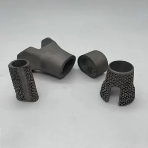
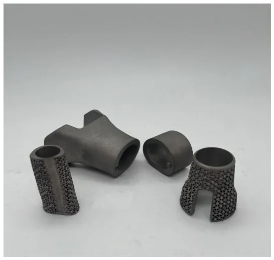
Using the CT scan images, a 3D model of the patient’s bone is created. After planning and determining the patient’s treatment plan, custom prostheses are designed for the patient.
The prosthesis was created using a 3D printer and, post-processing, sanitizing, and sterilization with gamma radiation, then it was provided to the doctor.
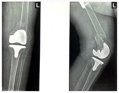
The result of the surgery was very satisfactory, and the patient’s bone lesion was completely reconstructed. She was able to walk the day after the surgery.