Anterior Cervical Discectomy and Fusion Interbody Cages
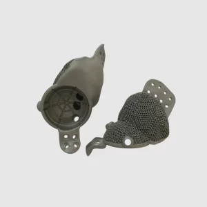
ACDF interbody cages for spinal fusion serve as a space holder between the vertebras, manufactured in different sizes in the case of spinal trauma, disc degeneration or degradation, and deformity correction. These cages use modern 3D printing technology and biocompatible medical-grade materials to improve patient contentment with surgical results.
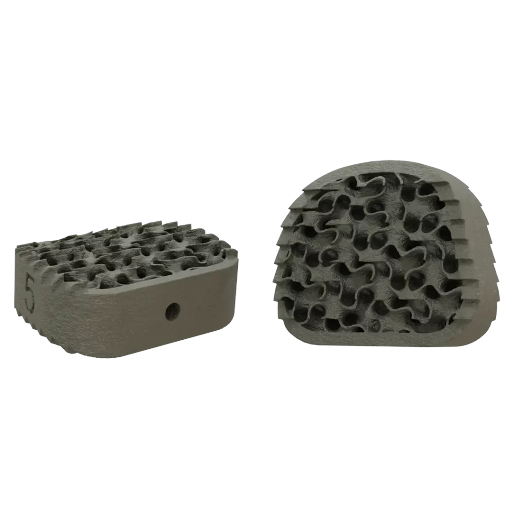
Product Overview
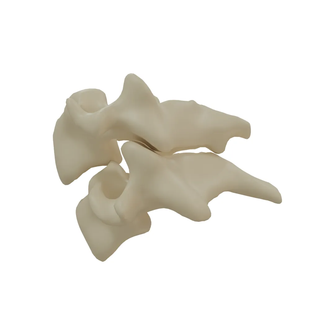
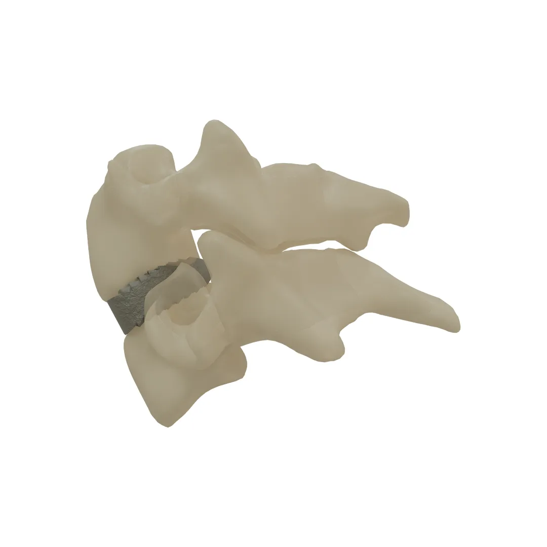
Product Overview
ACDF cages are available in sizes 5,6,7,8,9 to ensure optimal compliance with the patient’s anatomy and the physician’s treatment plan.
When these cages with aforethought porosity, teeth, and engineered slope design are created using biocompatible medical-grade materials and revolutionary 3D printing technology, the chances of a successful surgical outcome are significantly boosted.
These cages are the best option for patients with cervical trauma, disc degeneration, and deformity correction.
Workflow
Cage Design
Considering the disc location, and soft tissue, biomechanical engineers create these cervical cages in different sizes.
Product Manufacturing
3D printing technology is used to manufacture the cage with titanium.
Surgery
A set of sterilized cages in a few sizes is given to the physician. An employee of our company may serve in the operating room to help with cage placement.
Specifications
Together, surgeons and the engineering team have designed the cage’s form and functionalities based on the clinical assessment of patients and the intended course of treatment.
Our cages are designed with optimized porous structures to enhance cell growth, proliferation, adhesion, and optimal conditions for fusion to replace cervical intervertebral discs and to fuse the adjacent vertebral surfaces and to facilitate osseointegration.
Specially aligned teeth on superior and inferior surfaces ensure good primary stability and reduce the risk of cage migration as flared ends.
It is an optimized design to withstand loads applied to the spine. It is the best size for the optimum stress distribution pattern.
Designed in various sizes to adapt to different anatomy and designed to accommodate bone graft placement. Also, it creates a porous structure similar to the elastic modulus of bone to prevent stress shielding.
Specifications
Together, surgeons and the engineering team have designed the cage’s form and functionalities based on the clinical assessment of patients and the intended course of treatment.
benefits
Lorem ipsum dolor sit amet, it amet, consectetur adipiscing elit. Ut elit tellus, luctus nec ullamcorper mattis, pulvinar dapibus leo.Lorem , luctus nec ullamcorper mattis, pulvinar dapibus leo.
Lorem ipsum dolor sit amet, consectetur adipiscing elit. Ut elit tellus, luctus nec ullamcorper mattis, pulvinar dapibus leo.
- Lorem kjsae ismcp ipsum
- Lorem ipsur sit amet
- fast and hard
- easy and flex
Cases report
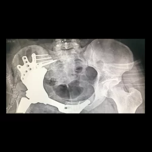
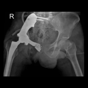
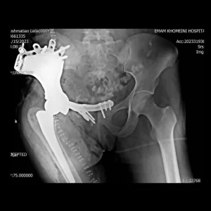
The patient is a 31-year-old man diagnosed with pelvic osteosarcoma, who presented with...Read More
The patient is a 36-year-old man who has been diagnosed with pelvic sarcoma...Read More
The patient is a 22-year-old woman who has been affected by a Giant...Read More
The patient is a 49-year-old man who had previously undergone surgery and subsequently...Read More
Cases report
Our case studies illustrate the benefits that come from utilizing our distinctively developed equipment. They demonstrate the effectiveness of our treatment approaches and the ability of our professionals to create unique solutions for the patients. Examining our reports may help you gain a better understanding of the level of precision and attention to detail we put into each and every aspect of our work with cages.

