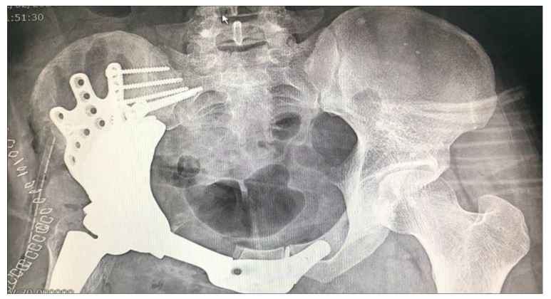
The patient is a 31-year-old man diagnosed with pelvic osteosarcoma, who presented with pain in the pelvic region.
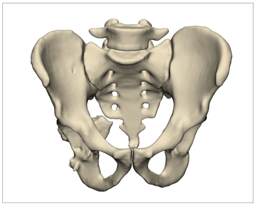
Pre-operative simulation and planning
Using MRI imaging, the locations of the cuts on the patient’s bone in the pubis and ilium were identified.
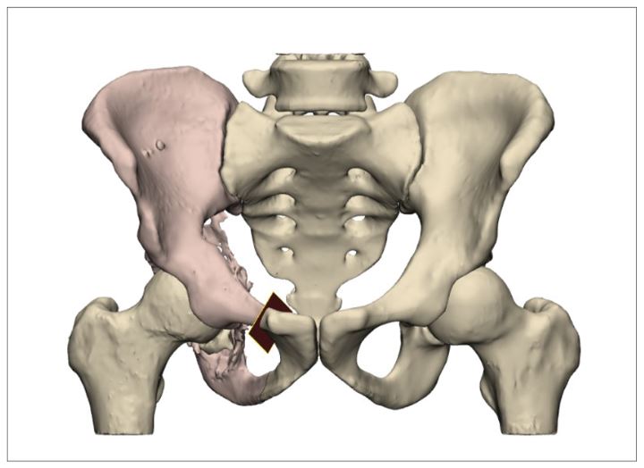
Based on the patient’s contralateral side, the overall geometry of the prosthesis was obtained. The version and inclination angles of the cup were also applied according to the patient’s healthy side.
Positions for 4.5 mm screws were designated at the connection of the prosthesis to the ilium. The fixation of the prosthesis on the pubis was performed on both the affected and healthy sides of the pelvis, with one 3.5 mm screw on the affected side, and one 4.5 mm screw along with the remaining 3.5 mm screws on the other side of the pelvis. Additionally, to ensure proper fixation, three 6.5 mm screw holes were created from inside the cup to secure the prosthesis to the wing of the ilium. A porous structure was created at the prosthesis-bone interfaces to promote bone tissue formation.
The part was manufactured using a 3D printer and, after post-processing, cleaning, and sterilization with gamma radiation, it was provided to the physician.
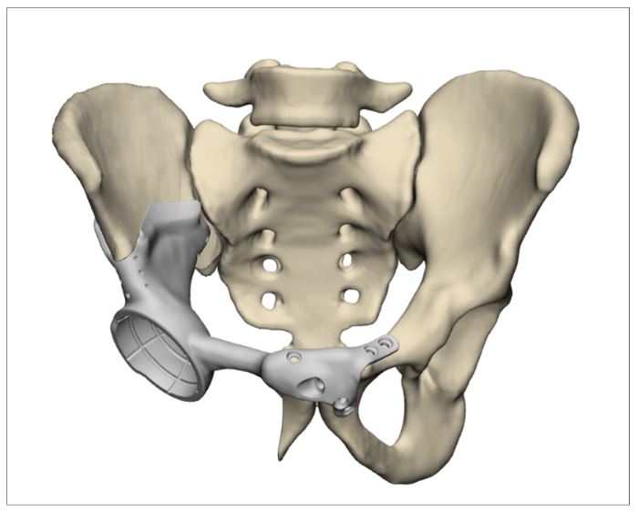
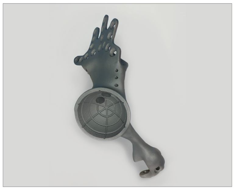
Tumor images
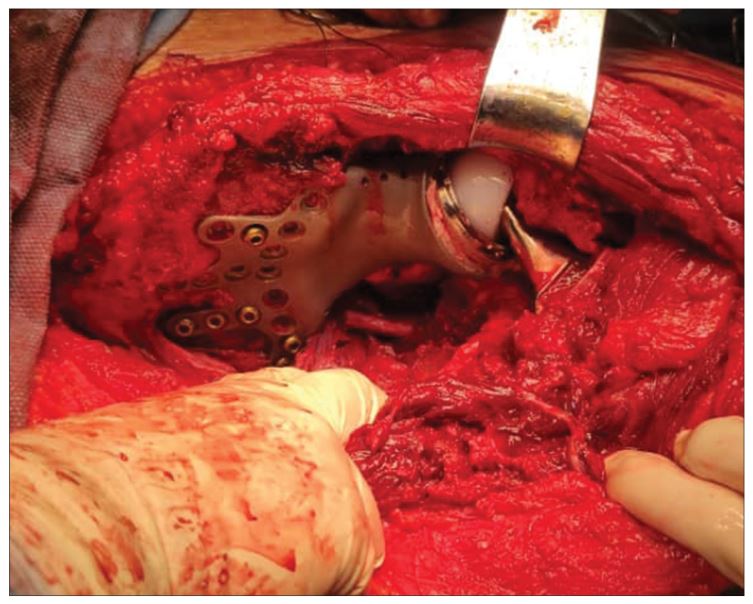
During the surgery, the guide designed based on the patient’s anatomy was first placed on the pubis bone, and the incision was made according to the geometry of the prosthesis, followed by tumor removal.
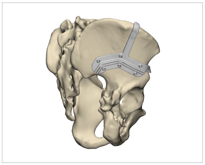
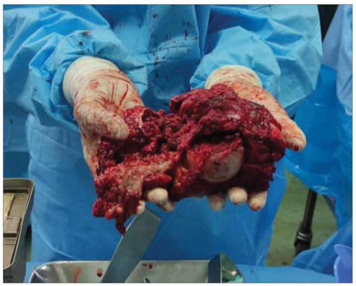
Surgical images
Based on preoperative simulation, the prosthesis was placed in the desired location.
Postoperative images
As shown in the postoperative image, the prosthesis fixation was achieved through a 6.5 mm screw inside the cup, as well as 3.5 mm and 4.5 mm screws in the pubis and ilium. The prosthesis was successfully positioned in the designated location.
