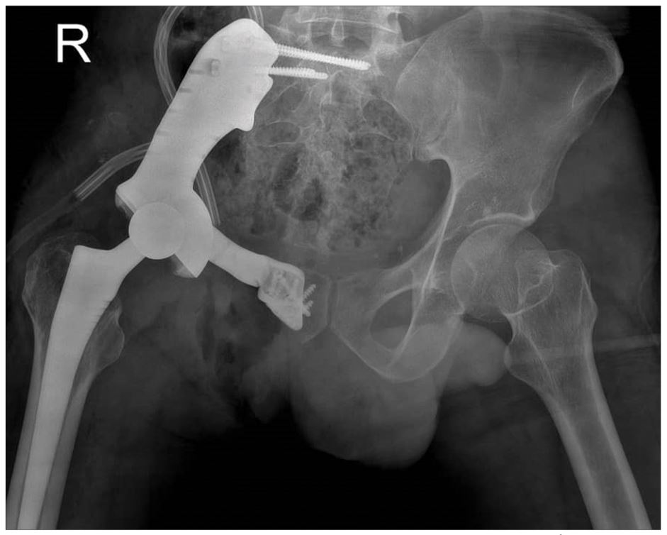
The patient is a 36-year-old man who has been diagnosed with pelvic sarcoma and presented with pain in the pelvic region.
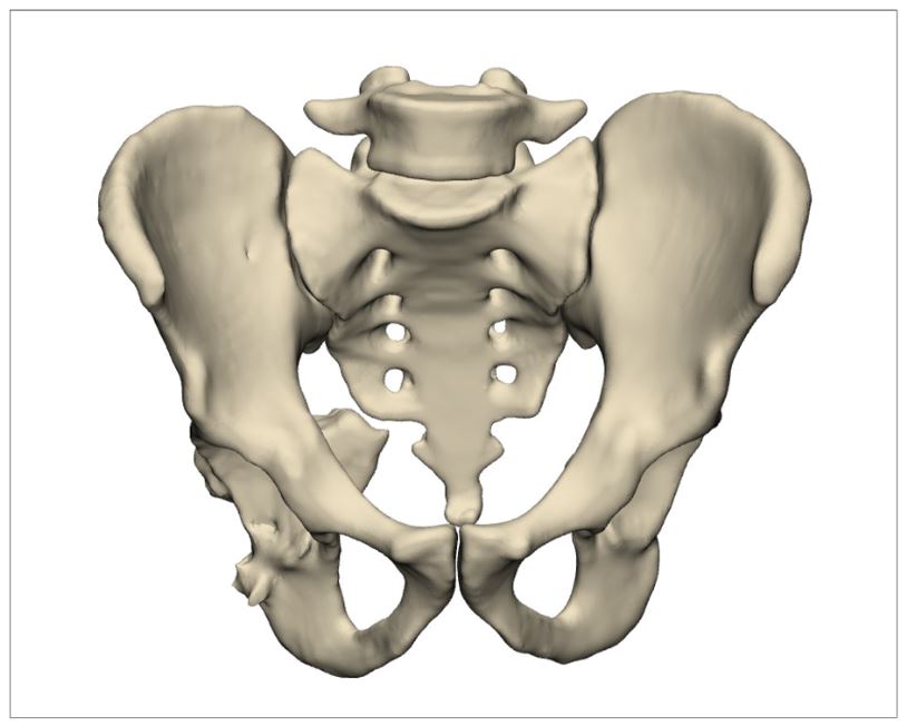
Pre-operative simulation and planning
Using MRI imaging, the locations of the cuts on the patient’s bone in the pubis were identified. Due to the involvement of the pelvis extending to the SI joint, reconstruction and expansion of the tumor to this area were necessary.
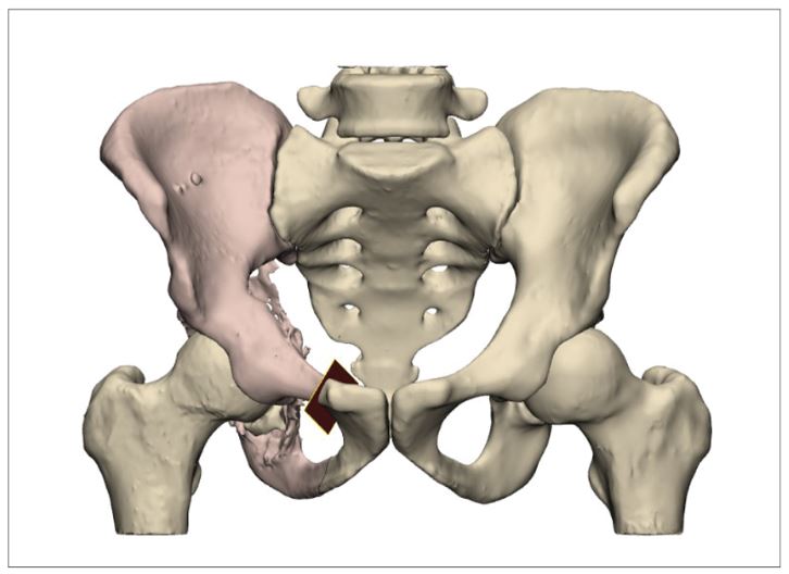
Based on the patient’s contralateral side, the overall geometry of the prosthesis was obtained. The version and inclination angles of the cup were also applied according to the patient’s healthy side. Positions for 4.5 mm screws were designated at the connection of the prosthesis to the sacrum, and 3.5 mm screws were placed at the connection of the prosthesis to the pubis. A porous structure was created at the prosthesis-bone interfaces to promote bone tissue formation.
The part was manufactured using a 3D printer and, after post-processing, cleaning, and sterilization with gamma radiation, it was provided to the physician.
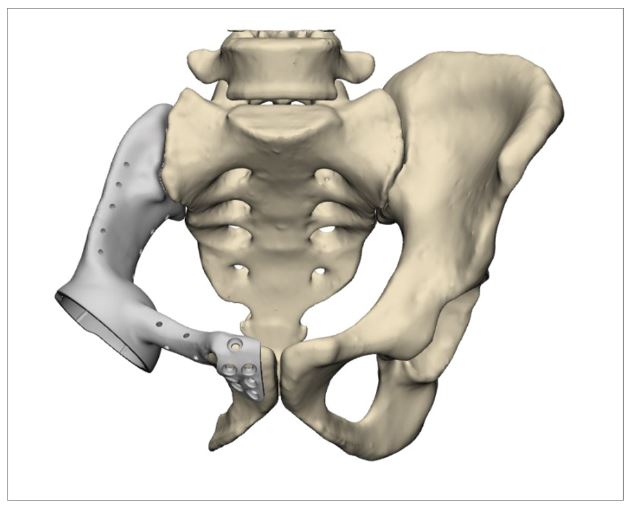
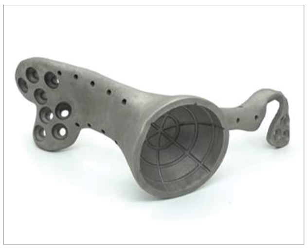
Tumor images
During the surgery, the guide designed based on the patient’s anatomy was first placed on the pubis bone, and the incision was made according to the geometry of the prosthesis.
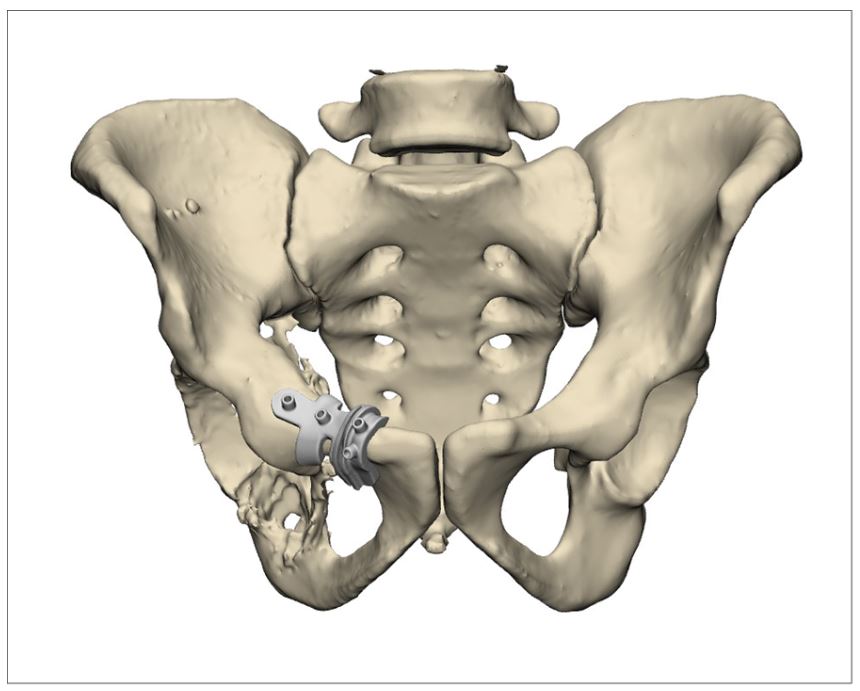
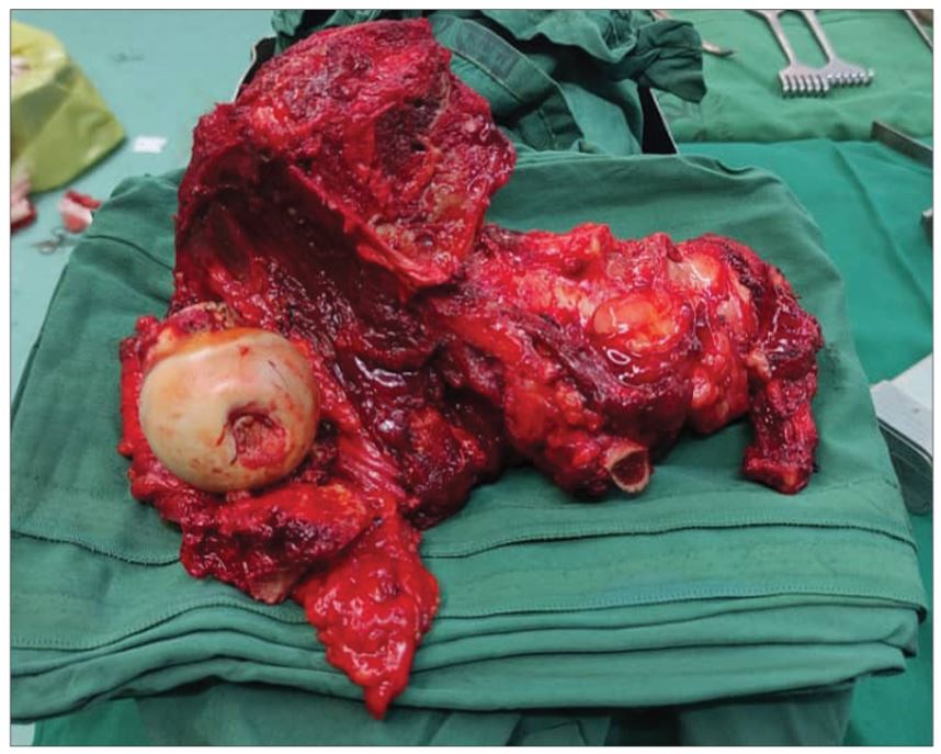
Images after surgery
The prosthesis was positioned on the bone according to the planning, and as shown in the postoperative image, the fixation of the prosthesis was achieved through the designated screw holes.
