3D Assessment Report
Using advanced imaging technology and carefully researched indices, our report provides surgeons with the information they need to make informed decisions and achieve optimal surgical outcomes.

Product Overview

Product Overview
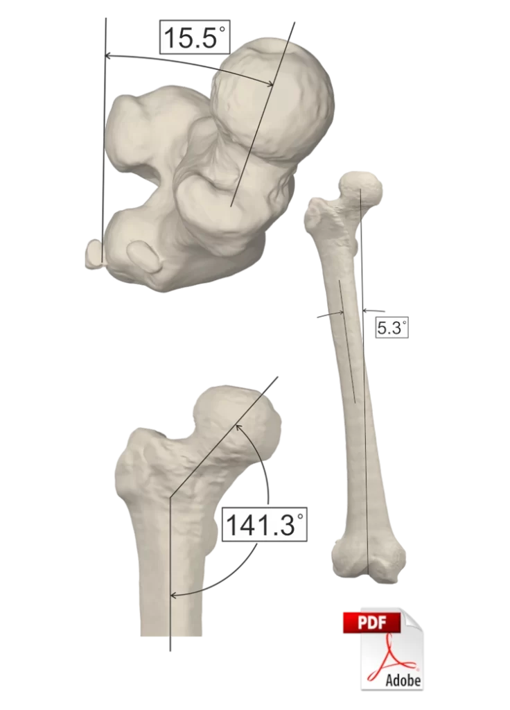
The 3D alignment assessment report provides a precise and advanced evaluation of the lower limb. Unlike simple AP radiographs, it takes into account the complex anatomy of the patient’s lower limb, including the femoral anteversion, tibial rotation, and TT-TG. Conventional radiographs can’t accurately determine these measurements, which is why having a complete 3D model of the patient’s lower limb based on CT scans and X-ray images is essential.
As a result of our 3D alignment assessment report, the surgeon receives a comprehensive evaluation of the patient’s lower limb alignment, along with detailed measurements and indices. In this way, every patient receives personalized care based on their unique anatomy, which allows for a more precise and accurate surgical approach. Moreover, our report includes a 3D model of the limb in PDF format that gives the surgeon a deeper understanding of the patient’s anatomy and streamlines the planning process.
Workflow
CT Scan
CT-Scan will be used to reconstruct an accurate 3D model of the patient.
XRay Imaging
XRay images will be used in order to transform supine CT Scan into a weight bearing image
Report
A comprehensive report of 3D assessment of patient's lower limb will be generated
Specifications
These are some distinctive characteristics that differentiate our 3D assessment reports from conventional AP radiographic images.
The 3D bone geometry generated from CT imaging is transformed into a weight-bearing model using X-ray radiographs, enabling 3D measurements of indices involving both the tibia and femur.
Our assessment reports provide a comprehensive evaluation of the limb by extracting 32 indices from literature and research articles, presenting a holistic assessment for the surgeon. The major advantage of our reports is that all necessary measurements are included, making it a one-stop-shop for the surgeon’s needs.
Having access to the 3D geometry of bones, our measurements are not affected by the viewing angle, and each bone can be placed in the desired orientation for accurate and repeatable results for each index measurement
Specifications
These are some distinctive characteristics that differentiate our 3D assessment reports from conventional AP radiographic images.
Benefits
Conventionally, a simple radiograph of three joints is usually requested as a preliminary step, and if the surgeon suspects any additional deformities, they may or may not ask for additional imaging to measure factors such as anteversion, TT-TG, or tibial torsion. However, with the 3D assessment approach, all measurements are obtained in a comprehensive manner, allowing for a complete evaluation of the patient’s anatomy. This approach ensures that all indices outside the normal range are brought to the surgeon’s attention, thereby minimizing the likelihood of missing any abnormality. The 3D assessment report provides a more holistic and accurate picture of the lower limb, making it an essential tool for surgeons dealing with complex anatomies or severe distortions. By allowing for a complete evaluation of the limb’s 3D geometry and providing measurements for all relevant indices, our approach ensures a more precise and effective surgical plan.
In cases where there is severe distortion of the anatomy, traditional 2D imaging may not be sufficient to accurately assess and study the affected area. By using 3D assessment, each bone can be evaluated as a whole and in its proper orientation, allowing for more precise measurements and a better understanding of the overall anatomy. In addition, many landmark and index definitions that may be used in 2D imaging may no longer be applicable in severely distorted anatomies. 3D assessment allows for a more comprehensive evaluation that takes into account all aspects of the anatomy, including any abnormalities or distortions that may be present.
Cases report
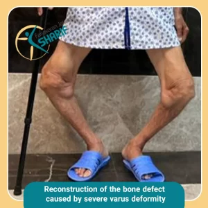
An 87-year-old male patient with severe genu varum (Varus-bow-legged) and significant bone defects...
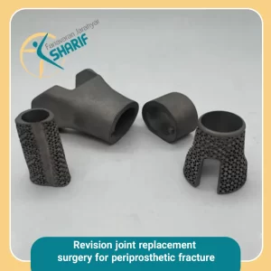
The patient is an 86-year-old woman who presented to the doctor with a...
A 45-year-old male patient who had suffered a fracture and comminution of the...
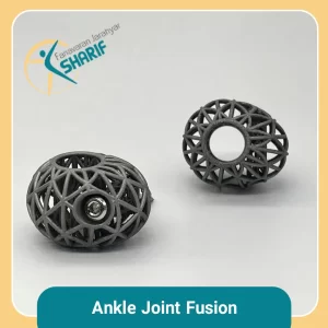
A 56-year-old male patient with Charcot joint due to diabetes had a portion...
Cases report
Our case reports highlight successful outcomes achieved through our custom-made implants. They showcase the effectiveness of our treatment plans and the expertise of our team in creating tailored solutions for each patient.
By reviewing these reports, you can gain a better understanding of the level of care and precision that we bring to every aspect of our work with patient specific implants.

